
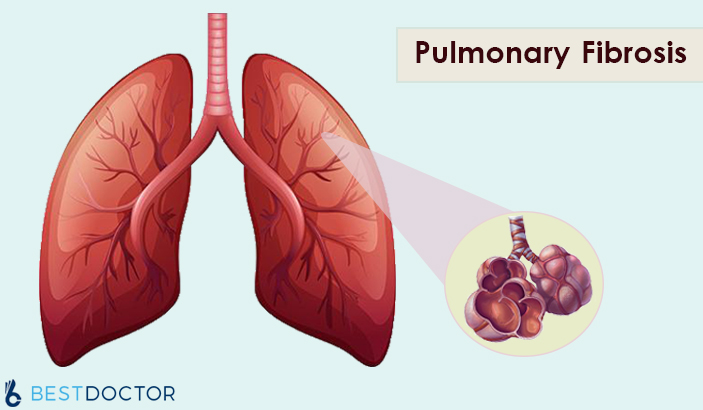
What Is Pulmonary Or Residual Fibrosis?
Pulmonary or residual fibrosis is a lung disease that occurs when lung tissue becomes damaged and scarred. This thickened, stiff tissue makes it more difficult for the lungs to work properly.
The scarring associated with pulmonary fibrosis can be caused by a multitude of factors. But in most cases, doctors can’t pinpoint what’s causing the problem. When a cause can’t be found, the condition is termed idiopathic pulmonary fibrosis.
Signs and Symptoms of Pulmonary Fibrosis may Include:
- Shortness of breath (dyspnea)
- A dry cough
- Fatigue
- Unexplained weight loss
- Aching muscles and joints
- Widening and rounding of the tips of the fingers or toes (clubbing)
Residual Fibrosis Causes
Occupational and environmental factors
Long-term exposure to a number of toxins and pollutants can damage your lungs. These include:
- Silica dust
- Asbestos fibers
- Hard metal dusts
- Coal dust
- Grain dust
- Bird and animal droppings
Radiation Treatments
Some people who receive radiation therapy for lung or breast cancer show signs of lung damage months or sometimes years after the initial treatment. The severity of the damage may depend on:
- How much of the lung was exposed to radiation
- The total amount of radiation administered
- Whether chemotherapy also was used
- The presence of underlying lung disease
Medications
Many Drugs can Damage the Lungs, Especially Medications such as:
- Chemotherapy drugs: Drugs designed to kill cancer cells, such as methotrexate (Trexall, Otrexup, others) and cyclophosphamide, can also damage lung tissue.
- Heart medications: Some drugs used to treat irregular heartbeats, such as amiodarone (Cordarone, Nexterone, Pacerone), may harm lung tissue.
- Some antibiotics: Antibiotics such as nitrofurantoin (Macrobid, Macrodantin, others) or ethambutol can cause lung damage.
- Anti-inflammatory drugs: Certain anti-inflammatory drugs such as rituximab (Rituxan) or sulfasalazine (Azulfidine) can cause lung damage.
Medical Conditions
Lung Damage can also Result from a number of Conditions, Including:
- Dermatomyositis
- Polymyositis
- Mixed connective tissue disease
- Systemic lupus erythematosus
- Rheumatoid arthritis
- Sarcoidosis
- Scleroderma
- Pneumonia
Many substances and conditions can lead to residual fibrosis. Even so, in most cases, the cause is never found. Pulmonary fibrosis with no known cause is called idiopathic pulmonary fibrosis.
Risk Factors
Factors that make you more Susceptible to Pulmonary Fibrosis Include:
- Age: Although pulmonary fibrosis has been diagnosed in children and infants, the disorder is much more likely to affect middle-aged and older adults.
- Sex: Idiopathic pulmonary fibrosis is more likely to affect men than women.
- Smoking: Far more smokers and former smokers develop pulmonary fibrosis than do people who have never smoked. Pulmonary fibrosis can occur in patients with emphysema.
- Certain occupations: There is an increased risk of developing pulmonary fibrosis if one works in mining, farming or construction or if exposed to pollutants known to damage lungs.
- Cancer treatments: Having radiation treatments to the chest or using certain chemotherapy drugs can increase the risk of pulmonary fibrosis.
- Genetic factors: Some types of pulmonary fibrosis run in families, and genetic factors may be a component.
Complications
Complications of Pulmonary or Residual Fibrosis may Include:
- High blood pressure in your lungs (pulmonary hypertension).
- Right-sided heart failure (corpulmonale).
- Respiratory failure.
- Lung cancer.
- Lung complications. As pulmonary fibrosis progresses, it may lead to complications such as blood clots in the lungs, a collapsed lung or lung infections.
Diagnosis
Imaging Tests
- Chest X-ray. A chest X-ray shows images of your chest. This may show the scar tissue typical of pulmonary fibrosis, and it may be useful for monitoring the course of the illness and treatment.
- Computerized tomography (CT) scan. CT scanners use a computer to combine X-ray images taken from many different angles to produce cross-sectional images of internal structures in the body. A high-resolution CT scan can be particularly helpful in determining the extent of lung damage caused by pulmonary fibrosis. Also, some kinds of fibrosis have characteristic patterns.
- Echocardiogram. An echocardiogram uses sound waves to visualize the heart. It can produce still images of your heart’s structures, as well as videos that show how your heart is functioning. This test can evaluate the amount of pressure occurring in the right side of your heart.
- Lung function tests
- Pulmonary function testing. Several types of pulmonary function tests may be conducted. In a test called spirometry, you exhale quickly and forcefully through a tube connected to a machine. The machine measures how much air your lungs can hold and how quickly you can move air in and out of your lungs. Other tests may be conducted to measure your lung volumes and diffusing capacity.
- Pulse oximetry. This simple test uses a small device placed on one of your fingers to measure the oxygen saturation in your blood. Oximetry can serve as a way to monitor the course of the disease.
- Exercise stress test. An exercise test on a treadmill or stationary bike may be used to monitor your lung function when you’re active.
- Arterial blood gas test. In this test, your doctor tests a sample of your blood, usually taken from an artery in your wrist. The oxygen and carbon dioxide levels in the sample are then measured.
- Tissue sample (biopsy)
If other tests haven’t diagnosed the condition, then doctors may need to remove a small amount of lung tissue (biopsy). The biopsy is then examined in a laboratory to diagnose pulmonary fibrosis or rule out other conditions. The tissue sample may be obtained in one of these ways:- Bronchoscopy
During bronchoscopy, your doctor may conduct an additional procedure called bronchoalveolar lavage. In this procedure, your doctor injects salt water through a bronchoscope into a section of your lung, and then immediately suctions it out. The solution that’s withdrawn contains cells from your air sacs. - Surgical biopsy. Although a surgical biopsy is more invasive and has potential complications, it may be the only way to obtain a large enough tissue sample to make an accurate diagnosis. This procedure may be done as a minimally invasive surgery, called video-assisted thoracoscopic surgery (VATS), or as an open surgery (thoracotomy).
- Blood tests to evaluate the liver and kidney function, and to test for and rule out other conditions.
- Bronchoscopy
Treatment
The lung scarring that occurs in pulmonary fibrosis can’t be reversed, and no current treatment has proved effective in stopping the progression of the disease. Some treatments may improve symptoms temporarily or slow the disease’s progression. Others may help improve quality of life. Doctors will evaluate the severity of your condition to determine the most appropriate treatment for your condition.
Medications
Newer medications are recommended now, including pirfenidone (Esbriet) and nintedanib (Ofev). These medications may help slow the progression of idiopathic pulmonary fibrosis. Both medications have been approved by the Food and Drug Administration (FDA). Additional medications and new formulations of these medications are being developed but have not yet been FDA approved.
Nintedanib can cause side effects such as diarrhea and nausea. Side effects of pirfenidone include rash, nausea, and diarrhea.
Oxygen Therapy
Using oxygen can’t stop lung damage, but it can:
-
-
- Make breathing and exercise easier
- Prevent or lessen complications from low blood oxygen levels
- Reduce blood pressure in the right side of your heart
- Improve your sleep and sense of well-being
-
Pulmonary Rehabilitation
Pulmonary rehabilitation can help you manage your symptoms and improve your daily functioning. Pulmonary rehabilitation programs focus on:
-
-
- Physical exercise to improve your endurance
- Breathing techniques that may improve lung efficiency
- Nutritional counseling
- Counseling and support
- Education about the condition
-
Lung Transplant
Lung transplantation may be an option for people with pulmonary fibrosis. Having a lung transplant can improve your quality of life and allow you to live a longer life. However, a lung transplant can involve complications such as rejection and infection.
Lifestyle and Home Remedies
Being actively involved in your own treatment and staying as healthy as possible are essential to living with pulmonary or residual fibrosis. For that reason, it’s important to:
-
- Stop smoking.
- Eat well.
- Get moving. Regular exercise can help you maintain your lung function and manage the stress.
- Get vaccinated.
- Follow your treatment plan.
[cl-review quote=”Medically Reviewed By” author=”Dr. Kaushal M. Bhavsar (MBBS, MD)” occupation=”Assistant Professor in Pulmonary Medicine, GMERS Medical College, Ahmedabad” avatar_image=”1325″ source=”url:https%3A%2F%2Fwww.linkedin.com%2Fin%2Fdr-kaushal-bhavsar-a8137355%2F|target:_blank”]


Why Indian Doctors Are Chosen By The UK NHS?
Norovirus – Symptoms, Treatment & Precautions
Understanding Normal Resting Heart Rate in Men and Women
5 Things to Learn about a Dentist before Choosing Them